Knee Muscle Anatomy Mri / Imaging Of Athletic Injuries Of Knee Ligaments And Menisci Sports Imaging Series Radiology. There is a flat area of tendon originating from the knee. The common peroneal nerve typically courses downward within abundant fat posterior to the short head of the biceps femoris muscle and superficial to the lateral head of the gastrocnemius muscle, but. The medial and lateral gastrocnemius tendons together with the soleus muscle form the calf. The smaller bone that runs alongside the tibia (fibula) and the kneecap (patella) are the other bones that make the knee joint. Superiorly, it extends to the level of the crossing of the biceps femoris tendon, and remains superficial to fcl in this location.10
The quadriceps muscles provide strength and power with knee extension (straightening). These motions of the knee allow the body to perform such important movements as walking, running, kicking, and jumping. Atlas of knee mri anatomy. Magnetic resonance imaging (mri) is a noninvasive test used to diagnose medical conditions. The medial and lateral gastrocnemius tendons together with the soleus muscle form the calf.
Knee Mri Anatomy from anatomia.wum.edu.pl When a muscle has different orientations of the tendons it means that there are different patterns of edema possible depending on the tendon injured. Anatomy basic knee mri checklist. Position the patient in supine position with feet pointing towards the magnet (feet first supine) position the patient over the spine coil and place the body coils over the thighs (anterior superior iliac spine down to knee joints) securely tighten the body coil using straps. Superiorly, it extends to the level of the crossing of the biceps femoris tendon, and remains superficial to fcl in this location.10 Stanford msk mri atlas has served over 1,000,000 pages to users in over 100 countries. These muscles work in groups to flex, extend and stabilize the knee joint. Related posts of knee muscle anatomy mri muscle anatomy coloring sheets. It is considered a vestigial muscle, and can be used as a tendon graft in reconstructive orthopedic surgery.
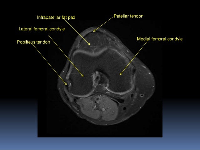
T2w axial fat sat 1.
Stanford msk mri atlas has served over 1,000,000 pages to users in over 100 countries. Abnormal anatomy with normal signal, i.e. Prescribe sagittal plane off axial images with line parallel to bony glenoid. The common peroneal nerve typically courses downward within abundant fat posterior to the short head of the biceps femoris muscle and superficial to the lateral head of the gastrocnemius muscle, but. Radiology imaging medical imaging subscapularis muscle shoulder anatomy bicep tendonitis mri brain shoulder rehab rotator cuff tear anatomy this. The medial and lateral gastrocnemius tendons together with the soleus muscle form the calf. Doctors may recommend a knee mri if a patient experiences the following(3): There is a flat area of tendon originating from the knee. In this presentation mri anatomy biceps femoris muscle. Anatomy basic knee mri checklist. This mri knee cross sectional anatomy tool is absolutely free to use. Plantaris acts weakly to plantar flex the foot and flex the knee. Plantaris can have variable size, but in most cases is difficult to demonstrate on routine mri studies.
Mri imaging palnes for pectoralis muscle. Song, uc san francisco msiv gillian lieberman md. Three conventional mri planes that are utilized to evaluate the knee include sagittal (oblique), coronal, and transaxial planes. There is a flat area of tendon originating from the knee. Medical images from an mri allow medical professionals to distinguish body tissues, including the meniscus (shock absorbers in the knee), cartilage, tendons, and ligaments.
The Knee Mri Atlas Of Anatomy In Medical Imagery from www.imaios.com The images may also help physicians to distinguish normal, healthy tissues from dead tissues(2). Mri imaging palnes for pectoralis muscle. This long muscle flexes the knee. Knee anatomy the orthopedic sports medicine institute in they act like strong ropes to connect bones. Medical images from an mri allow medical professionals to distinguish body tissues, including the meniscus (shock absorbers in the knee), cartilage, tendons, and ligaments. Anatomy arthrogram anatomy basic shoulder mri. Articular surface of patella and femur, condyle, epicondyle and muscles (popliteus, sartorius, gastrocnemius, semimembranous with tendos.) the images obtained were exported to jpeg from dicom data stored on the pacs (picture archiving and communicating system). Related posts of knee muscle anatomy mri muscle anatomy coloring sheets.
T2w axial fat sat 1.
Plantaris acts weakly to plantar flex the foot and flex the knee. T2w axial fat sat 1. The muscles of the knee include the quadriceps, hamstrings, and the muscles of the calf. Intensity corresponds to a pathologic lesion. Atlas of knee mri anatomy. Knee anatomy the orthopedic sports medicine institute in they act like strong ropes to connect bones. Anatomy basic knee mri checklist. Superiorly, it extends to the level of the crossing of the biceps femoris tendon, and remains superficial to fcl in this location.10 To realign the anterior cruciate ligament parallel with the sagittal imaging plane. The common peroneal nerve typically courses downward within abundant fat posterior to the short head of the biceps femoris muscle and superficial to the lateral head of the gastrocnemius muscle, but. There are various muscles that control movement ligaments that give stability special cartilage to absorb pressure and various other structures to ensure smooth pain. In this presentation mri anatomy biceps femoris muscle. In conclusion, we describe the normal mri anatomy of the distal biceps femoris and the relationship of this muscle with the common peroneal nerve.
Mri knee anatomy knee sagittal anatomy free cross sectional anatomy mri knee mri diagnostic imaging : A tendon connects the muscle to the bone. This mri knee cross sectional anatomy tool is absolutely free to use. Song, uc san francisco msiv gillian lieberman md. Anatomy arthrogram anatomy basic shoulder mri.
Mri Knee Joint Anatomy from image.slidesharecdn.com The smaller bone that runs alongside the tibia (fibula) and the kneecap (patella) are the other bones that make the knee joint. Song, uc san francisco msiv gillian lieberman md. The knee joint is most significantly affected by two major muscle groups: When a muscle has different orientations of the tendons it means that there are different patterns of edema possible depending on the tendon injured. Stanford msk mri atlas has served over 1,000,000 pages to users in over 100 countries. Use the mouse scroll wheel to move the images up and down alternatively use the tiny arrows (>>) on both side of the image to move the images. Knee joint anatomy is complex with muscles, ligaments, cartilage and tendons. Plantaris can have variable size, but in most cases is difficult to demonstrate on routine mri studies.
The muscles that affect the knee's movement run along the thigh and calf.
Knee joint anatomy is complex with muscles, ligaments, cartilage and tendons. The quadriceps muscles provide strength and power with knee extension (straightening). The images may also help physicians to distinguish normal, healthy tissues from dead tissues(2). Extending along the anterior surface of the thigh are the four. Doctors may recommend a knee mri if a patient experiences the following(3): The smaller bone that runs alongside the tibia (fibula) and the kneecap (patella) are the other bones that make the knee joint. Use the mouse scroll wheel to move the images up and down alternatively use the tiny arrows (>>) on both side of the image to move the images. This mri knee cross sectional anatomy tool is absolutely free to use. The common peroneal nerve typically courses downward within abundant fat posterior to the short head of the biceps femoris muscle and superficial to the lateral head of the gastrocnemius muscle, but. Abnormal anatomy with normal signal, i.e. Familiarity with the normal gastrocnemius tendon mri features, as well as the spectrum of mri findings with different types of pathology that may involve these structures is relevant for accurate interpretation of knee mri. Articular surface of patella and femur, condyle, epicondyle and muscles (popliteus, sartorius, gastrocnemius, semimembranous with tendos.) the images obtained were exported to jpeg from dicom data stored on the pacs (picture archiving and communicating system). Position the patient in supine position with feet pointing towards the magnet (feet first supine) position the patient over the spine coil and place the body coils over the thighs (anterior superior iliac spine down to knee joints) securely tighten the body coil using straps.
Share this post
0 Response to "Knee Muscle Anatomy Mri / Imaging Of Athletic Injuries Of Knee Ligaments And Menisci Sports Imaging Series Radiology"


0 Response to "Knee Muscle Anatomy Mri / Imaging Of Athletic Injuries Of Knee Ligaments And Menisci Sports Imaging Series Radiology"
Post a Comment